Searchable clinical interpretation library

Laboratory & clinical investigations

Choose a test to understand what it measures, its normal reference values, visual interpretation, common pitfalls and findings that require urgent escalation.

Use your laboratory rangeReference intervals, assays and critical-value policies vary. Interpret results with the patient, trend, medicines and local guidance.

Genuine clinical and microscopy examples

Real-life laboratory interpretation atlas

Synthetic report pictures removed. These de-identified teaching specimens show actual morphology; numeric results must still be interpreted using the patient’s own laboratory report.
Haematology

Read the FBC, indices, reticulocytes and blood film together

Film morphology directs the differential but rarely establishes the cause alone.

Normal blood smear containing erythrocytes, platelets and a neutrophil1Uniform red cells2Mature neutrophil3Platelets

Normal peripheral blood film

A real Wright-stained film showing broadly uniform red cells, a mature neutrophil and small platelets.

Look for
Cell size, central pallor, variation in shape/size, white-cell maturity and platelet estimate.
Clinical next step
Use as morphology context only; normal morphology does not exclude quantitative cytopenia or early disease.
Echinaceapallida, CC BY-SA 4.0 · original and licence ↗
Peripheral blood smear showing severe microcytic hypochromic iron deficiency anaemia1Small red cells2Large central pallor3Marked size/shape variation

Severe microcytic, hypochromic iron-deficiency pattern

This patient’s MCV was 55 fL. The film shows marked microcytosis, hypochromia, anisocytosis and poikilocytosis.

Look for
Low Hb with low MCV/MCH; confirm iron deficiency with ferritin and transferrin saturation while accounting for inflammation.
Clinical next step
Find the cause—blood loss, diet, malabsorption or increased demand. Severe symptoms, instability or active bleeding require urgent management.
Ed Uthman, CC BY 2.0 · original and licence ↗
Peripheral blood film from a patient with megaloblastic anaemia showing a hypersegmented neutrophil1Hypersegmented neutrophil2Macro-ovalocytes

Megaloblastic macrocytic pattern

A genuine film from megaloblastic anaemia. Hypersegmented neutrophils and macro-ovalocytes support impaired DNA synthesis.

Look for
High MCV, B12/folate status, reticulocyte response, medicines, alcohol/liver disease, thyroid disease and marrow disorders.
Clinical next step
Do not give folate alone until important B12 deficiency is addressed; neurological features require prompt treatment and specialist advice.
Ed Uthman, CC BY 2.0 · original and licence ↗
Peripheral blood film showing fragmented red blood cells or schistocytes1Fragmented RBC2Helmet-shaped cell3Cell fragments

Microangiopathic haemolysis—schistocytes

Fragmented red cells support mechanical or microangiopathic destruction in the right clinical setting.

Look for
Anaemia, thrombocytopenia, raised LDH/unconjugated bilirubin, low haptoglobin, reticulocytosis and organ injury.
Clinical next step
Schistocytes with thrombocytopenia, neurological/renal injury or suspected TTP/DIC require immediate haematology escalation.
Osaro Erhabor, CC0 · original and licence ↗
Peripheral blood smear from a patient with leukaemia showing circulating blasts1Large immature blast2High nuclear-to-cytoplasm ratio

Acute leukaemia—circulating blasts

A genuine leukaemic blood film with immature blast cells. Automated counts may flag rather than classify them.

Look for
Blasts, cytopenias or very high WBC, constitutional symptoms, bleeding, infection and tumour-lysis features.
Clinical next step
Discuss immediately with haematology; avoid delay when there is leukostasis, DIC, sepsis or tumour lysis.
Ajay Kumar Chaurasiya, CC BY-SA 4.0 · original and licence ↗
Giemsa-stained blood smear showing sickled red cells and target cells1Sickled red cell2Target cell

Sickle-cell disease film

Giemsa-stained film showing characteristic sickle forms and target cells.

Look for
Clinical crisis phenotype, Hb, reticulocytes, haemolysis markers, oxygenation and prior haemoglobin analysis.
Clinical next step
The film supports morphology; diagnosis and genotype require haemoglobin analysis. Treat acute chest syndrome, stroke or sepsis as emergencies.
Keith Chambers, CC BY-SA 3.0 · original and licence ↗
CSF

Appearance + opening pressure + cells + protein + paired glucose + microbiology

No single picture or number reliably separates every cause. Prior antibiotics and disease timing change the pattern.

Real cerebrospinal fluid specimen with purulent cloudy appearance1Cloudy / purulent appearance

Purulent CSF in acute bacterial meningitis

A visibly turbid CSF specimen. Gross appearance can suggest a heavy inflammatory burden but clear CSF does not exclude meningitis.

Look for
Raised opening pressure, neutrophilic pleocytosis, high protein and low CSF:serum glucose ratio; Gram stain, culture and PCR refine cause.
Clinical next step
If bacterial meningitis is suspected, give antibiotics within the time-critical pathway—do not delay for imaging or LP.
Whein, CC BY 3.0 DE · original and licence ↗
Stained CSF microscopy showing numerous neutrophils in purulent meningitis1Numerous neutrophils2Inflammatory cellular background

Neutrophilic CSF pleocytosis

Pappenheim-stained CSF with numerous neutrophils, consistent with purulent meningitis in context.

Look for
Neutrophils commonly suggest bacterial infection, but early viral and partially treated disease can overlap.
Clinical next step
Send differential count, protein, glucose with paired serum glucose, culture/Gram stain and targeted PCR. Discuss unusual patterns with microbiology.
Jensflorian, CC BY-SA 3.0 · original and licence ↗
India ink or nigrosin preparation of CSF showing encapsulated Cryptococcus1Encapsulated yeast2Clear halo

Cryptococcal microscopy

A real CSF negative-stain preparation showing encapsulated yeast. Modern cryptococcal antigen testing is more sensitive than India ink.

Look for
Raised opening pressure, lymphocytic or low-cell CSF, high protein, low glucose and positive cryptococcal antigen/culture.
Clinical next step
Urgent infection/HIV specialist management; measure and actively manage raised intracranial pressure.
Meditechnika, CC BY-SA 4.0 · original and licence ↗
CT image of intracerebral and intraventricular haemorrhage used to explain that CSF xanthochromia follows a bleeding pathway1Acute intracranial blood2Intraventricular extension

Subarachnoid haemorrhage—xanthochromia is a laboratory diagnosis

This CT demonstrates intracranial blood. When the SAH pathway requires LP after negative imaging, xanthochromia is assessed by the local laboratory—visual tube colour alone is unreliable.

Look for
RBC count pattern, spectrophotometric bilirubin/xanthochromia and timing from ictus; distinguish traumatic tap using the full local pathway.
Clinical next step
Thunderclap headache remains an emergency. Follow the current CT/LP protocol and urgently discuss positive findings with stroke/neurosurgery.
Yadav et al., CC BY 2.0 · original and licence ↗
Oncology and haematological malignancy

Use tumour markers and proteins for specific questions—not as a screening panel

Interpret the assay, trend and pre-test probability, then confirm disease anatomically and histologically where appropriate.

Serum protein electrophoresis showing a monoclonal M spike in multiple myeloma1Narrow monoclonal M peak2Gamma region

Monoclonal gammopathy—M spike

A protein electrophoresis trace showing a narrow monoclonal peak. The size alone does not separate MGUS from myeloma.

Look for
SPEP/immunofixation, serum free-light-chain ratio, Hb, calcium, creatinine, symptoms and imaging.
Clinical next step
AKI, hypercalcaemia, severe anaemia, hyperviscosity or cord compression requires urgent haematology assessment.
Steven Fruitsmaak, CC BY-SA 3.0 · original and licence ↗
Bone marrow smear showing acute myeloid leukaemia with blasts and Auer rods1Myeloblast2Auer rod3Prominent nucleolus

AML marrow—blasts and Auer rods

A marrow smear from AML with maturation, showing blasts and Auer rods. Morphology is followed by flow cytometry, cytogenetics and molecular classification.

Look for
Cytopenias or leukocytosis, blasts, DIC (especially suspected APL), infection, bleeding and tumour lysis.
Clinical next step
Immediate haematology discussion; suspected APL with coagulopathy is a time-critical emergency.
AFIP/PEIR, public-domain US government work · original and licence ↗
PET CT showing metabolically active metastatic cervical lymph node1FDG-avid nodal disease2Anatomical CT correlation

Tumour-marker interpretation

A real PET/CT with FDG-avid nodal metastasis illustrates why biochemical markers must be correlated with imaging and tissue diagnosis.

Look for
Indication-specific marker: PSA, CA-125, AFP, β-hCG, CEA or CA19-9; note benign causes, assay and trend.
Clinical next step
A normal marker never excludes cancer. Use NICE suspected-cancer pathways and urgent escalation for cord compression, SVC obstruction or tumour lysis.
Akira Kouchiyama, CC BY-SA 3.0 · original and licence ↗

Educational image atlas: these images demonstrate genuine morphology, not diagnostic-quality patient reports. Always review the original specimen/report, units, reference interval, analyser comments, clinical context and local urgent-result policy.

New enhanced section

Interpretation of medical images

Chest X-ray • CT • MRI • PET/CT • ultrasound
Use a fixed system, compare prior imaging and read the radiologist’s report. These examples teach recognition; they do not replace diagnostic image review.
1ConfirmPatient, date/time, modality, body part, projection/sequence, contrast and clinical question.
2QualityCoverage, rotation, inspiration/exposure, motion and artefact. Poor quality changes confidence.
3SearchUse the modality-specific checklist below; inspect every region, not only the obvious lesion.
4ConcludeDescribe finding, location, severity, complications, comparison and urgent action.
AP chest radiograph showing right upper lobe airspace shadowing from pneumonia1Right upper-lobe airspace opacity2Compare both hila3Check pleural angles

Chest X-ray—right upper-lobe pneumonia

A real AP chest radiograph showing right upper-lobe shadowing. Consolidation is a pattern, not an organism.

Look for
RIPE quality, then A–E: airway, breathing/lungs/pleura, circulation/mediastinum, diaphragm, everything else. Seek air bronchograms, silhouette loss and complications.
Clinical next step
Correlate with physiology and infection syndrome. Follow-up imaging is indicated for selected patients according to local/NICE guidance.
CDC, public domain · original and licence ↗
Chest radiograph showing a large right spontaneous pneumothorax with visible pleural line1Visceral pleural line2No peripheral lung markings3Assess mediastinal shift

Chest X-ray—large right pneumothorax

A genuine chest radiograph showing a large right pneumothorax. Tension is a clinical diagnosis supported by imaging when time permits.

Look for
Pleural line, absent peripheral markings, lung collapse, deep sulcus on supine film and mediastinal shift.
Clinical next step
Unstable suspected tension pneumothorax requires immediate decompression—do not delay for imaging.
James Heilman MD, CC BY 3.0 · original and licence ↗
Non-contrast CT brain showing intracerebral and intraventricular haemorrhage1Hyperdense acute blood2Intraventricular extension3Assess mass effect / hydrocephalus

CT brain—intracerebral haemorrhage

On non-contrast CT, acute blood is typically hyperdense. Inspect every slice and window; this single teaching frame cannot show the full extent.

Look for
Blood, extra-axial collections, early ischaemic change, mass effect, midline shift, ventricles/cisterns, skull and sinuses.
Clinical next step
Immediate stroke/neurosurgical pathway; reverse anticoagulation when indicated and manage physiology according to current guidance.
Glitzy queen00, public domain · original and licence ↗
CT pulmonary angiography images showing saddle pulmonary embolus and bilateral thrombus burden1Central filling defect2Saddle embolus3Assess RV strain

CT pulmonary angiography—saddle embolus

CTPA demonstrates contrast-opacified pulmonary arteries with intraluminal filling defects.

Look for
Embolus level/burden, RV:LV ratio and septal bowing, reflux into IVC/hepatic veins, infarction and alternative diagnoses.
Clinical next step
Risk-stratify using haemodynamics, biomarkers and RV findings. Shock or deterioration requires immediate senior/PERT/critical-care escalation.
Thrombosis Journal open-access case, CC BY · original and licence ↗
MRI brain showing deep cerebellar haemorrhage1Cerebellar lesion2Posterior fossa

MRI brain—cerebellar haemorrhage

MRI signal depends on sequence and blood age. DWI/ADC is central to acute ischaemia; FLAIR, T1/T2 and susceptibility answer different questions.

Look for
Confirm sequence and plane; compare DWI with ADC, inspect FLAIR/T2, SWI/GRE for blood, vessels, mass effect and enhancement after contrast.
Clinical next step
Interpret with onset time and neurological examination. Posterior fossa lesions can deteriorate rapidly despite small size.
Andrewmeyerson derivative, CC BY-SA · original and licence ↗
PET CT images showing FDG avid cervical lymph node metastasis1High FDG uptake2CT anatomical lesion3Review whole-body distribution

PET/CT—FDG-avid nodal metastasis

PET displays metabolism; CT supplies anatomy. Uptake is not synonymous with malignancy because inflammation and physiological activity also take up FDG.

Look for
Pattern and intensity of uptake, anatomical correlate, physiological sites, treatment timing, blood glucose and prior study.
Clinical next step
Use for the specific staging/response question and multidisciplinary interpretation; biopsy remains necessary when tissue diagnosis changes management.
Akira Kouchiyama, CC BY-SA 3.0 · original and licence ↗
Ultrasound image showing a normal gallbladder without stones or wall thickening1Anechoic gallbladder lumen2Thin wall3No posterior shadowing stone

Abdominal ultrasound—normal gallbladder view

A genuine normal gallbladder ultrasound image. Ultrasound is dynamic and operator-dependent; a still frame never replaces the full examination.

Look for
Gallstones and shadowing, wall thickness, pericholecystic fluid, sonographic Murphy sign and common bile duct calibre.
Clinical next step
Escalate suspected biliary sepsis or obstruction; correlate with LFTs, inflammatory markers and the formal report.
Ptrump16, CC BY-SA 4.0 · original and licence ↗
Chest X-ray

RIPE quality → A–E review → compare prior. Never stop after the first abnormality.

CT

Confirm contrast phase and windows. Scroll systematically; one screenshot cannot exclude disease.

MRI

Know the sequence. Signal meaning changes between DWI/ADC, FLAIR, T1/T2 and susceptibility imaging.

PET/CT

Metabolic uptake needs anatomical and clinical correlation; infection/inflammation can be FDG-avid.

Ultrasound

Orientation, probe position, dynamic signs and Doppler matter; static images are incomplete.

Safety: If imaging and the patient conflict, reassess the patient, verify the study and contact radiology. Act immediately on time-critical findings such as tension pneumothorax, major haemorrhage, large-vessel occlusion, aortic catastrophe, massive PE, cord compression or perforation.

PICTURE-GUIDED CLINICAL PROCEDURE PATHWAYS

Bedside procedures

Select a procedure for a stepwise safety, technique, samples and aftercare pathway.

STAND-ALONE BEDSIDE PROCEDURE PATHWAY

Lumbar puncture

Sterile CSF sampling with opening pressure and correctly prioritised tubes
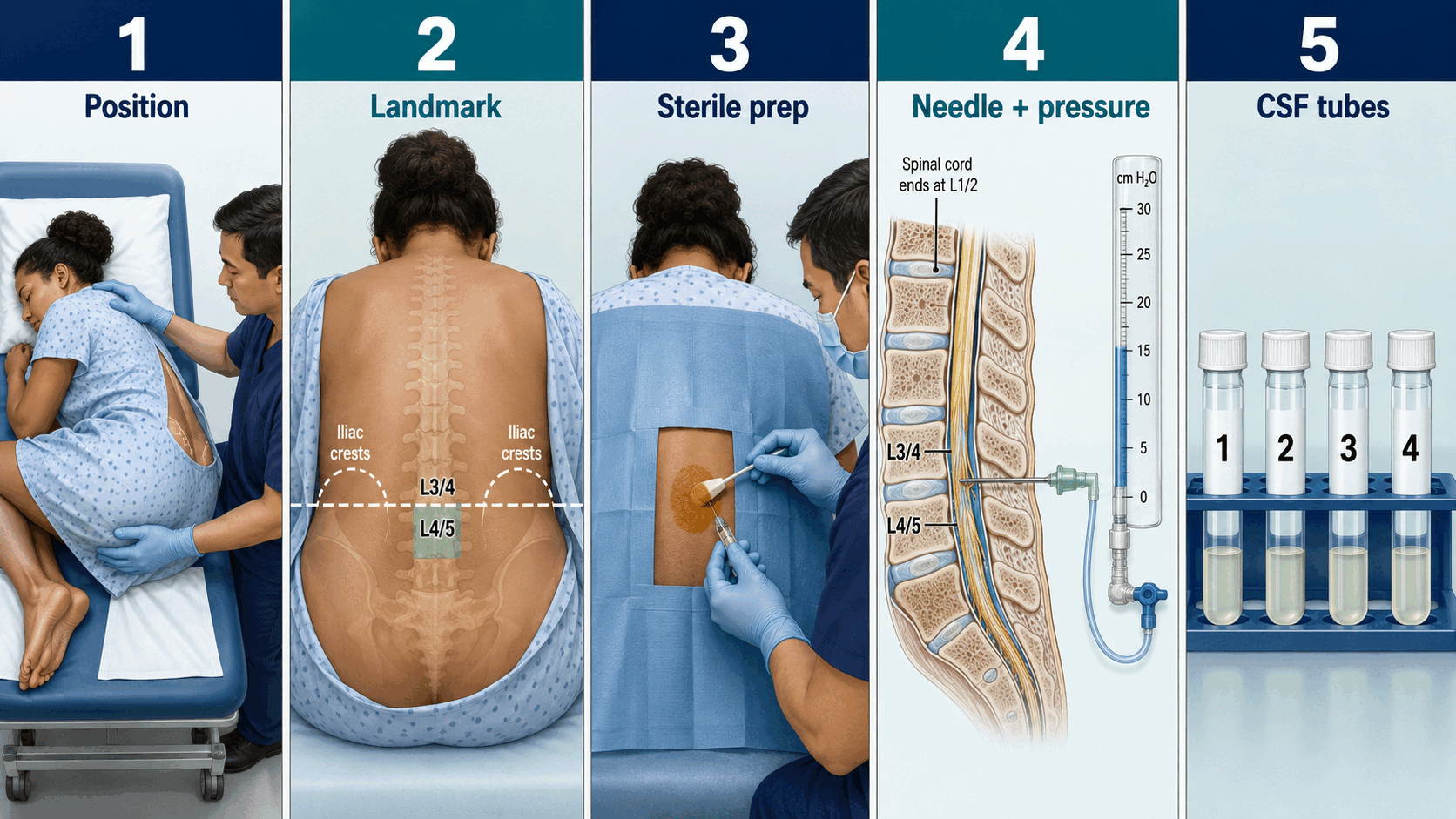
Adult lumbar puncture positioning, landmarking, sterile preparation, spinal needle and pressure measurement, and sequential CSF tubes
1Position2Landmark3Sterile prep4Needle + pressure5CSF tubes

Indications

  • Suspected CNS infection/inflammation after time-critical treatment
  • Subarachnoid haemorrhage when the pathway requires CSF
  • Pressure disorders, malignancy and specialist neurological diagnosis

Stop / escalate

  • Mass lesion/obstructive hydrocephalus or clinical herniation risk
  • Infection at puncture site, major uncorrected bleeding risk or unsafe anticoagulation
  • Cardiorespiratory instability—stabilise first; do not delay antibiotics/aciclovir

Illustrated step sequence

1Consent, neurological exam, platelets/coagulation/antithrombotics and imaging decision
2Lateral decubitus for valid opening pressure; identify L3/4 or L4/5 below the cord
3Full asepsis, local anaesthetic and atraumatic spinal needle where possible
4Measure pressure with legs relaxed; collect sequential labelled tubes without aspiration
5Document level, needle, attempts, pressure, appearance, tests and complications

Samples / technical essentials

  • Cells/differential, protein and glucose with paired serum glucose
  • Gram stain/culture and syndrome-specific PCR
  • Xanthochromia, oligoclonal bands with paired serum, cytology/flow or TB/fungal tests only when indicated

Aftercare and complications

  • Observe neurology, puncture site and clinical stability
  • Post-dural puncture headache: analgesia/hydration advice; anaesthetic review for persistent severe orthostatic headache
  • Urgent assessment for new deficit, sphincter disturbance, severe back/radicular pain, fever or bleeding

Supervised clinician education. Use the current local checklist, competency standard, consent policy, infection-control procedure and escalation route. This pathway does not certify procedural competence.

50 investigations • each includes reference values

Screen anaemia, infection/inflammation, cytopenias and haematological disease; always interpret components together and compare with baseline/trend.

Normal reference values

Illustrative adult guide • confirm against the patient’s report

HaemoglobinMen 130–180; women 115–165 g/LLOWNORMAL / EXPECTEDHIGH
WBC4.0–11.0 ×10⁹/LLOWNORMAL / EXPECTEDHIGH
Platelets150–400 ×10⁹/LLOWNORMAL / EXPECTEDHIGH
MCV80–100 fLLOWNORMAL / EXPECTEDHIGH
Ranges vary with laboratory, analyser, biological sex, age, pregnancy, units and clinical context.

How to interpret

  • Hb low: classify by MCV, reticulocytes and clinical context; assess bleeding, haematinic deficiency, haemolysis, renal disease and marrow disease.
  • Neutrophilia may reflect infection, inflammation, steroids or stress; neutropenia severity, duration, medicines and sepsis risk determine urgency.
  • Platelets: confirm unexpected thrombocytopenia on repeat/sample film; consider consumption, drugs, liver/splenic disease, immune and marrow causes.

Common pitfalls

  • Reference ranges vary by laboratory, age, sex and pregnancy.
  • Haemoconcentration or dilution can mislead; a normal total WBC does not exclude infection.
  • Review the blood film when counts are unexplained, severe or discordant.

Urgent findings

  • Neutropenic sepsis
  • Severe symptomatic anaemia or active bleeding
  • Very low platelets with bleeding, schistocytes or suspected TTP/DIC
Open specialist guidance ↗